
Single Photon Emitters
The NV single emitter series features fluorescent diamond containing very low amounts of NV centers per particles. These products are geared toward sensing applications, where the unique spin properties of the NV- center are exploited for use in signaling and sensing. Sizes available include 10nm and 40nm particles. Applications areas include, but are not limited to:
Quantum optics and quantum computing
Single photon emitter applications
Neuron firing detection
Single cell tracking and sensing
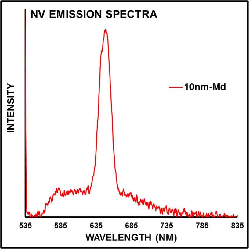
Figure 1: Fluorescent emission spectra for 10nm-Md suspension in DI water at approximately 1mg/mL concentration. 45mW 532nm laser excitation (Coherent Sapphire). Ocean Optics HR2000 spectrometer with 500msec integration time. Water Raman indicated by black arrow.
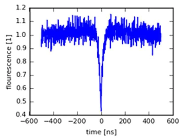
Figure 3: g(2) autocorrelation function of 8nm particle exhibiting spin coherence consistent with NV- presence. Courtesy T. Oeckinghaus, U. Stuttgart.
Combined HRTEM, AFM, and DLS confirms the presence of particles 10nm in size and below. Shapes are irregular, with some particles exhibiting flat, elongated shapes. Some particles exhibit an amorphous patches of sp2 carbon even after extensive oxidation.
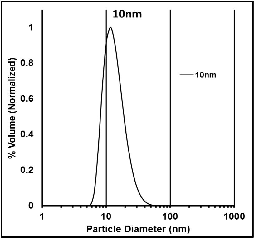
Figure 5: DLS size distribution of 10nm particles in DI water. Measured with Malvern Zetasizer Nano ZS (Malvern Instruments Ltd. UK)
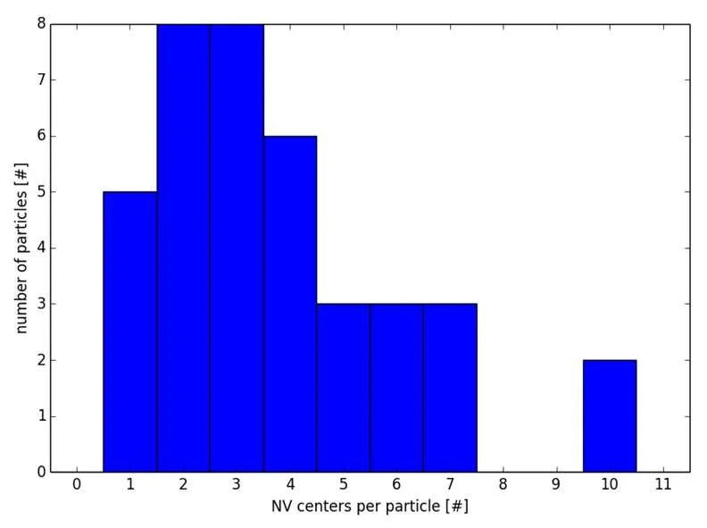
Figure 7: Distribution of NV centers per particle for 40nm-Lw as determined from AFM-Confocal single particle characterization. Courtesy T. Oeckinghaus, U. Stuttgart.
The 40nm single emitter series is the most popular product in use for quantum optics research groups. Single particle characterization determined the average number of emitters (NV centers) per particle to range from 1-4 on average. The NV⁻/NV⁰ ratio was determined to be ~0.7.
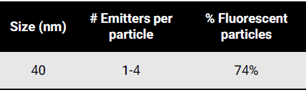
Table 2: Summary of single particle characterization of 40nm-Lw particles using AFM-Confocal microscopy setup. Courtesy T. Oeckinghaus, U. Stuttgart.
ULTRASMALL : 10-13nm
The level of fluorescence and the population of fluorescent diamond particles in the sub-15nm range is generally very low. These particles are not suitable for direct cellular imaging, but will require users equipped with highly advanced optical setups to use effectively. Therefore, these particles are more suitable for users interested in single emitter applications. The quality of NV⁻ centers in these particles is relatively low due to large lattice distortion and damage induced from milling from larger sizes. Single particle fluorescence characterization confirmed the presence of active NV⁻ centers, and determined that the approximate NV⁻/NV⁰ ratio was ~0.6, so fluorescing particles contain ~60% NV⁰ and ~40% NV⁻.

Table 1: Summary of single particle characterization of 10nm-Md particles using AFM-Confocal microscopy setup. Courtesy T. Oeckinghaus, U. Stuttgart.
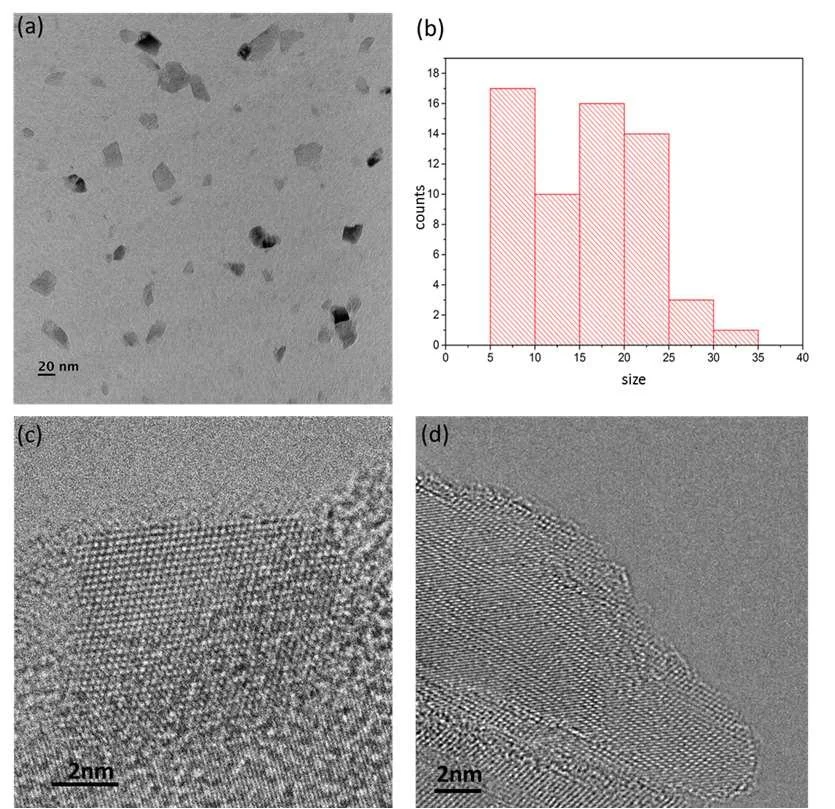
Figure 2: High Resolution TEM (HRTEM) images and size distribution of 10nm-Md particles. Courtesy S. Chang, U. Arizona.
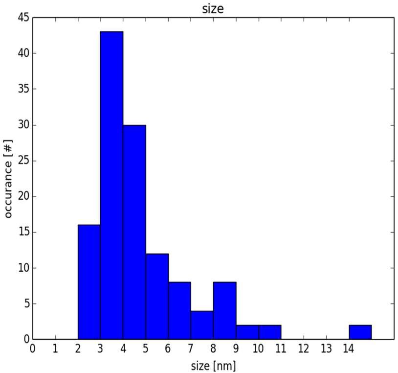
Figure 4: AFM confocal size (height) distribution of 10nm-Md particles. Courtesy T. Oeckinghaus, U. Stuttgart.
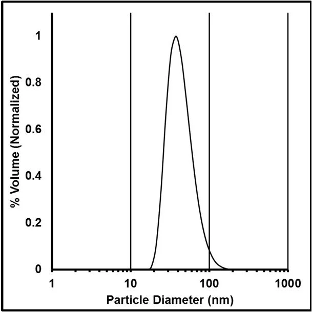
Figure 6: DLS size distribution of 40nm particles in DI water. Measured with Malvern Zetasizer Nano ZS (Malvern Instruments Ltd. UK)
Procedure for dispersion of ND-NV single particles individually over glass using spin coating.
Quite often nanodiamond particles containing NV centers (ND-NV) need to be dispersed over glass for characterization or application purposes. Below is a tentative procedure developed by our collaborators from academia to disperse ND-NV single particles individually over glass using spin coating. NDs terminated with carboxylic groups provide a reasonable adherence to glass substrate. The procedure includes use of polyvinyl alcohol (PVA) for a better adherence.
The recommended procedure is as following:
For spin-coating use concentrations of ND-NV ~0.01-0.05 mg/ml (in the final-solution the concentration will be only 50% of that because of the PVA-solution)
Prepare the the PVA-solution 0.3% (w/w)
Mix 15 μl of diamond-solution and 15 μl of PVA-solution (vortex and 10 min sonification in a bath)
Use spin rate 3000 rpm for 20s (not very critical)
Make sure your glass-slides are well cleaned (a "plasma-cleaner" can be used)
If the slide is plasma-cleaned, 30 μl is enough to cover the whole slide (e.g. 22x22 mm)