We are proud to announce a program of lectures on cutting-edge nanodiamond and “quantum” macroscale diamond research.
|
“Tailoring the Surface of Nanodiamond for Applications in Biomedicine, Quantum Technology and Catalysis” May 2024 – (date tbd), Noon EST (+17:00 UTC) |
Below are the recorded videos of lectures (some with transcripts) given by experts in the field.
Past Lecture Series |
||
Speakers |
Abstracts |
Videos |
|
“Tuning a Quantum Nanoprobe: Advancements in Diamond Chemistry using Halogens, Low-Z precursors and Metal Oxides” April 18, 2024, Noon EDT (+16:00 UTC) Quantum sensing and quantum communication technologies are emergent fields and will transform detection and cyber security in the 21st century. Nitrogen vacancy centers or NV centers are fluorescent defects hosted in diamond and can be described as a quantum bit (qubit) capable of magnetometry, electrometry and thermometry. The field of diamond surface chemistry is ripe for exploration as quantum sensing and quantum communication become mature with diamond and other exotic materials that host atomic defects. High-pressure high-temperature (HPHT) nanodiamond (ND) is the most common nanoscale host of NV centers and are typically purified with aerobic oxidation to form an oxygen rich surface. Past work has determined the surface to be largely terminated with alcohols similar to 111-terminated bulk diamond. In this work, a combination of halogen, low-Z and metal oxide chemistry on ND will be discussed. The chemical analysis is completed with overlapping laboratory and X-ray synchrotron-based spectroscopies. Chemical modalities using bromine, boron, nitrogen and silanes will be summarized and the results will be impactful for researchers using ultra-hard materials for quantum sensing applications and interested in controlling the surface properties including surface dipole moment and electronic noise. |
||
|
“Multimodal Quantum Metrology in Living Systems Using Nitrogen-Vacancy Centres in Diamond Nanocrystals” April 4th, 2024, Noon EST (+16:00 UTC) Next-generation biological sensors and diagnostic tools require high sensitivity and spatial resolution to be able to identify emergent biological behaviour. Correlating multiple interdependent parameters at the nanoscale could help uncover details of cellular response to external perturbations. Temperature and viscosity are key parameters of interest that relate to cellular energetics and metabolism, morphological changes, cell division and active transport. Cells respond to temperature through viscoadaptation, and a change in viscosity may in turn affect the local temperature profile. In this talk, I will present our latest results on nanoscale quantum sensing in living human cancer cells. We use a diamond nanocrystal quantum sensor containing nitrogen-vacancy colour centres to extract information about the nanoscale thermal environment, the thermal and stochastic forces acting on the nanodiamond, and the properties of its nanoscale viscoelastic environment. We will also discuss opportunities in diamond-based quantum sensing for biomedical applications. |
||
 |
“Perspectives in Quantum Diamond Technologies” Feb 2nd 2024, Noon ET Optically active spin qubits in diamond have recently emerged as a candidate for a number of quantum technology areas, including quantum information processing and quantum sensing. The electron spin of the nitrogen-vacancy (NV) center can be optically polarized and readout. In addition, the electron spin of the NV center has a long coherence time under ambient conditions. These unique optical and spin properties make the NV spin qubit a unique system for quantum sensing. In this talk, we will introduce the new and rapidly developing field of nanoscale quantum sensing based on NV centers in diamond and potential applications for molecular imaging in life sciences. We will also discuss applications of NV centers in diamond nanoparticles for polarization of nuclear spins and the application of diamond-based hyperpolarization markers in ultrasensitive MRI. |
|
|
“Measuring T1 relaxation from FNDs in solution: methodology and motivation” Dec 13th 2023, 4:30 PM EST (+21:30 UTC) Fluorescent nanodiamonds (FNDs) have been exploited as sensitive quantum probes for nanoscale chemical and biological sensing applications, with the majority of demonstrations to date relying on the detection of single FNDs containing either single NVs or NV ensembles. This places significant limits on the measurement time, throughput and statistical significance of a measured result as there is usually marked inhomogeneity within FND samples. In this talk I will cover our recently published work on a complimentary measurement platform that can report the T1 spin relaxation time from a large ensemble of FNDs in solution. I will describe the apparatus in detail as well as an example sensing study used to compare this technique against existing methods. Finally, I will cover the niche that we believe this sensing modality can fill, as well as envisioned improvements. |
“Diamond Based Quantum Sensing for Free Radical Detection in Medical Samples“ |
|
|
“Diamond Based Quantum Sensing for Free Radical Detection in Medical Samples“ Nov 30th 2023, Noon ET (+17:00 UTC) Free radical generation plays a key role in many biological processes including cell signalling, immune responses or ageing. On the other hand an access in free radical production is usually associated with stress responses and pathological conditions. However, free radical generation is challenging to measure since they are small, occur in low concentrations and are short lived. I the last years we have established diamond-based quantum sensing as a tool to measure free radical generation at the nanoscale. More specifically, we use nanodiamonds containing ensembles of nitrogen vacancy centers and track their movement while they are moving through living cells. At the same we measure the magnetic noise by relaxometry. Since free electrons in radicals cause such a signal, this method allows us to measure the free radical generations in the immediate surrounding of the particles. In this presentation, I will give an update on our progress in this field and on bringing this technique to the medical field. More specifically, I will show how we can measure disease status as well as drug effectiveness in living cells. |
“Diamond Based Quantum Sensing for Free Radical Detection in Medical Samples“ |
|
 Nicholas Nunn, North Carolina State University/Adamas Nanotechnologies
|
“Magnetic Resonance and Optical Studies of HPHT Synthesized Diamond Microparticles With Controlled Nitrogen Content“ Oct 26th 2023, Noon ET (+16:00 UTC) Diamond particles hosting nitrogen vacancy centers is a platform material for potential use in emerging nano and microscale sensing technologies. These envisioned quantum sensors are based on optical manipulation and readout of NV centers in response to external environmental stimuli (e.g., temperature, strain, magnetic fields). The important prerequisites for such measurement protocols are NV centers with suitable spin properties such as long coherence and relaxation times. While such properties are readily achieved for bulk diamond crystals of suitable quality, the properties of diamond particles are often severely degraded by the presence of paramagnetic defects such as electronic spins associated with other nitrogen defects (P1 centers). We demonstrate the growth of microparticulate diamond with good crystal quality and controlled nitrogen content in the range of ca. 5-50 atomic ppm is possible. A resultant improvement in NV coherence times by approximately 3-fold is observed using pulsed electron paramagnetic resonance (EPR) across the range of nitrogen concentration studied. These results may provide a foundation for the production of the next generation of fluorescent diamond particles, which thus far have relied exclusively on particulate diamond with nitrogen contents in the range of ca. 100 atomic ppm. |
|
 |
“Is there substitutional nitrogen in nanodiamonds? Solving a puzzle by magnetic resonance“ May 25th 2023, Noon ET (+16:00 UTC) One of the most actual scientific and technological challenges associated with the use of nanodiamonds (NDs) consists in fabrication of NDs particles with sizes less than 100 nm carrying high density of triplet (S=1) nitrogen vacancy centers (NV–). Substitutional nitrogen atoms in neutral charge state with electron spin S=1/2 (N0/C/P1 centers) are among fundamental precursors of NV–. Electron paramagnetic resonance (EPR) provides unambiguous identification and quantification of P1. P1 EPR spectra in both mono- and poly-crystalline diamond samples are characterized by well-resolved hyperfine structure (HFS). On diminishing fluorescent diamond microcrystallites’ size, drastic changes in EPR spectra occur: significant decrease in the abundance of NV–; successive decrease in contribution of the initial HFS split P1 signal; increase in contribution of a broad singlet signal; appearance and consistent growth of new narrow singlet signal. EPR spectra of small NDs (both crushed microdiamonds and detonation nanodiamonds-DNDs) consists of two structureless Lorentzians. In contrast, increasing the average size of NDs from nanometers to micrometers by sintering of DNDs under pressure leads to restoration of typical P1 EPR signals with resolved HFS pattern. Surveying the entire set of experimental EPR data obtained on a huge variety of diamond particles from micro- to nanometer size-range, obvious questions arise: (i)What does really happen with paramagnetic substitutional nitrogen on nanonization? (ii)Does the nitrogen atom migrate and/or change its charge state? (iii)How many P1 centers do really exist in NDs and DNDs? Proper analysis and interpretation of magnetic resonance data, especially EPR ones, allow adequate answering all these questions. It was assumed that, upon crushing of diamond particles, density of P1 practically doesn’t change and all effects concerned with disappearance of P1 HFS pattern and appearance of intensive narrow singlet component, occur due to dipole-dipole and exchange interactions of P1 centers with surface paramagnetic defects formed upon nanonization. |
“Is there substitutional nitrogen in nanodiamonds? Solving a puzzle by magnetic resonance“ |
|
“Emerging Quantum Sensing Technologies: Interfacing coherent qubits with biological targets” March 23rd 2023, Noon ET (+16:00 UTC) Quantum metrology enables some of the world’s most sensitive measurements. When applied to biophysical systems, diamond-based quantum sensors have the potential to probe processes that cannot be accessed by conventional technologies. Examples of such processes range from cancer research to neuroscience to developmental biology. However, interfacing coherent qubit sensors with fragile biological target systems has remained an outstanding challenge that has severely limited applications. In this talk, I will discuss a novel approach that combines single-molecule biophysics technology with quantum engineering to interface intact biomolecules on a diamond quantum sensor without impacting qubit coherence and bio-functionality. In a second part, I will discuss our recent work on engineering highly coherent quantum sensors based on diamond nanocrystal. Such nanosensors can readily be taken up by cells and integrated into intact organisms. However, coherence in these nanocrystal sensors is limited by surface noise, which severely reduces the sensor’s sensitivity. In our work we developed a new approach to engineer spin coherence in core-shell nanostructures which leads to a 50-fold improvement in qubit sensitivity. Finally, potential future applications of quantum sensing to biophysical and diagnostics will be discussed. |
“Emerging Quantum Sensing Technologies: Interfacing coherent qubits with biological targets” |
|
|
“Probing Nanoscale Magnetic Phenomena Using Diamond Quantum Sensing Microscopy” January 19th 2023, Noon EST Diamond quantum sensing microscopy based on nitrogen vacancy (NV) centers has become a versatile tool to detect magnetic fields with an unprecedented combination of spatial resolution and magnetic sensitivity, opening up new frontiers in biological and condensed physics matter research. In this talk I will present two examples of using NV magnetometry in both scanning probe microscopy (SPM) and wide-field microscopy (WFM) geometries to probe nanoscale magnetic phenomena in different materials. |
“Probing Nanoscale Magnetic Phenomena Using Diamond Quantum Sensing Microscopy” |
|
|
“Novel NMR Quantum Sensors via Hyperpolarized Nuclei“ November 10 2022, Noon ET We report on new experiments that demonstrate the potential of hyperpolarized nuclear spins to serve as sensitive magnetometers and chemical sensors of their local environment. Our goals are to leverage this to create new forms of sub-micron-scale NMR detectors using hyperpolarized nanoparticles. We demonstrate this with 13C nuclei in diamond hyperpolarized through Nitrogen-Vacancy (NV) centers. The methodology leverages the exquisite spin lifetimes possible in diamond 13C nuclei under RF driving, wherein we observe lifetimes in excess of T2’=90s at room temperature. When exposed to a time-varying (AC) magnetic field, the nuclei undergo secondary precessions that carry an imprint of its frequency and amplitude. Hyperpolarization and continuous spin readout enable significant gains in sensitivity and resolution. Fourier-limited resolution is better than 100 mHz and single-shot sensitivity is better than 70pT at a bias field of 7T, amongst the best for quantum sensing at high-fields. Overall, this suggests interesting new opportunities for spin sensors possible via hyperpolarized, low-gamma, nuclei. |
||
|
“Imaging Cellular and Intracellular Components Using Quantum Diamond Microscopy“ October 20 2022, Noon ET The Nitrogen Vacancy (NV) centre in diamond is becoming an increasingly investigated probe in biology owing to its excellent photoluminescence properties and low cytotoxicity profile. Indeed, the sensitivity of NV photoluminescence to external factors that perturb the NV spin and/or charge state is being exploited as a sensing scheme. Coupled with existing fluorescence microscopy techniques readily available in biological laboratories it is now possible to map and track critical cellular events such as free radical generation, electrical activity and iron uptake and release by metalloproteins. In this talk I will present details of NV sensing schemes deployed using a minimally modified commercial fluorescent microscope that could be readily deployed in biology labs by non-experts. I will also report example uses of this instrumentation imaging cellular and intracellular components in mammalian cells and mitochondrial extracts.
|
“Imaging cellular and intracellular components using quantum diamond microscopy“ |
|
|
“High-Resolution Quantum Diamond Magnetic Imaging for Applications in
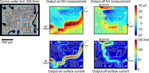
September 22 2022, 12 PM ET The quantum diamond microscope (QDM) is a technology used for magnetic imaging with high spatial resolution. Using a synthetic diamond sample with a surface layer of magnetically-sensitive NV centers to optically image the magnetic fields from external sources, a QDM offers micron-scale resolution over a few-mm field of view, high-reliability operation in ambient conditions, and high signal-to-noise readout of nano- and micro-scale magnetic sources. These attributes are enabling new scientific results that were previously impractical or impossible. In this talk I will present recent results applying NV magnetic microscopy to Sandia problems, including applications in electrical engineering, hardware security, condensed-matter physics, and quantum computing. |
||
|
|
“Surface Chemical Modification of Fluorescent Nanodiamonds Opens New Possibilities for NV Centers in the Measurement of Living Systems” June 23 2022, 9:00 AM ET, 2 PM London, 10 PM Tokyo How to measure biological changes quantitatively occurring inside cells is a significant issue in biology, which has remained unresolved for many years. Cells, with only a few tens of micrometers, are pretty small for such measurements. Moreover, each cell is further divided into smaller —micrometers to nanometers — compartments, each of which has its characteristic function, and the homeostasis of the cell system depends on the complex interactions among these small compartments. Therefore, to investigate intracellular systems quantitatively, novel technologies are a long-felt need for quantitative measurements of various parameters in the nanometer range. Recently, a fluorescent nanodiamond containing nitrogen-vacancy centers (NV centers) has attracted attention as a promising nanometer-scale measurement tool. An NV center is a lattice defect harboring a pair of triplet electrons, emitting 650-750 nm red fluorescence by photoexcitation with green light. The fluorescence intensity is known to be strictly conjugated with the spin state of the triplet electrons. Therefore, fluorescent nanodiamonds can be used for nanometer-scale measurement via microscopic fluorescence detection. Indeed, this idea is highly compatible with fluorescent bioimaging techniques such as cellular imaging and biological imaging and is expected to be applied to various biological fields, including cell biology, neuroscience, immunology, and so on. In this lecture, I would like to talk about how chemical modification of nanodiamond surfaces can extend the possibilities of microenvironment measurements of biological samples using nanodiamond NV centers. In particular, I’ll mention specific molecular and cellular measurements enabled by modifying nanodiamonds with hyperbranched polyglycerol and pH nanosensors realized by enriching a particular chemical group. |
“Surface Chemical Modification of Fluorescent Nanodiamonds Opens New Possibilities for NV Centers in the Measurement of Living Systems” |
|
Haksung Jung, Korea Research Institute of Standards and Science Keir C. Neuman, National Heart, lung, and Blood Institute, National Institutes of Health |
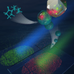
“Development of Fluorescent Nanodiamonds for Latent Fingerprint Detection” May 26th 2022, 11:00 AM ET Latent fingerprints (LFPs) are one of the most important forms of evidence in crime scenes due to the uniqueness and permanence of the friction ridges that comprise fingerprints. An efficient method to detect LFPs is therefore crucial in forensic science. However, traditional detection strategies are limited by low sensitivity, low contrast, high background, and complicated processing steps. Taking advantage of the remarkable optical and physical properties of fluorescent nanodiamonds (FNDs), we developed an approach that provides biocompatible, efficient, and low background latent fingerprint detection with polyvinylpyrrolidone (PVP) coated FNDs (FND@PVP). We demonstrate that PVP-coated FNDs are biocompatible at the cellular level, neither inhibiting proliferation nor eliciting an immune response. We then show that PVP coating enhances the physical adhesion of FNDs, resulting in efficient binding to latent fingerprint ridges due to the intrinsic amphiphilicity of PVP. Clear, well-defined ridge structures with first, second, and third-level LFP details are revealed within minutes by FND@PVP. The combination of this binding specificity and the remarkable optical and physicochemical properties of FND@PVP permits the detection of LPFs with high contrast, efficiency, selectivity, sensitivity, and reduced background interference. Our results demonstrate that FND@PVP nanoparticles have great potential for high-resolution imaging of LFPs. |
“Development of Fluorescent Nanodiamonds for Latent Fingerprint Detection” |
|
|
“Diamond Quantum Biosensors; Opportunities and Challenges” April 28th 2022, 4:30 ET Nanodiamonds are carbon-based nanomaterials that have attracted significant attention over the past decade. Fluorescent nanodiamonds (FNDs) provide a unique range of chemical, physical, and biological properties which have driven their application in the fields of drug delivery, tissue engineering, bioimaging and quantum biosensing. Recent studies exploiting the quantum properties of the nitrogen-vacancy (NV) centre in FNDs have demonstrated the detection of magnetic, electric, and temperature fields, as well as pH, redox and radical species. These quantum sensors therefore open up new and exciting opportunities to monitor biological activity at the nanoscale. In this talk, I will outline our efforts in applying FND probes to a range of biological problems. I will describe how these materials can be used to enhance to probe the intracellular temperature of living cells and monitor paramagnetic molecules in solution. I will discuss our recent work exploiting 2D NV imaging arrays for magnetic microscopy. In particular, I will show how these magnetic imaging techniques can be used to non-invasively map the magnetic properties of iron-oxide complexes in biological systems at the sub-cellular scale. I will also discuss the future possibilities of these technologies and how they might be applied to address significant and outstanding questions in biology. |
“Diamond quantum biosensors; opportunities and challenges” |
|
Professor Somnath Bhattacharyya, University of the Witwatersrand, South Africa
|
“Diamond-Based Hybrid Qubit System for Quantum Computation Technology“ February 17th 2022, Noon ET There is a surge in exploring the best qubit platform for building efficient quantum computers suitable for complex operations. A qubit is analogous to the binary bit seen in a conventional computer. We show the potential for a diamond-based hybrid quantum system combing electrical (superconducting) and optical (nitrogen-vacancy centers) techniques. Superconducting qubit technology typically involves the fabrication of various logic elements based on Josephson junctions using standard superconducting materials such as Al or Nb. Boron-doped superconducting diamonds are however attractive due to better resistance to external perturbations. Considering a recent discovery of triplet-spin-based superconductivity in nanocrystalline diamond films and that diamond presents well-established quantum states in the form of nitrogen-vacancy centers (NV-centers), diamond demonstrates a potential platform for the realization of all diamond-based quantum circuitry. We advance the understanding of superconducting order parameters in boron-doped nanodiamond films and as a new type of qubit system through the designing of the resonator and superconducting quantum interference devices. Quantum information can be exchanged between a superconducting diamond loop (with a phase-slip qubit junction) and an ensemble of spins (NV- centers) attached to a microwave line of the diamond nanowire that would allow us to initialize, control through microwave circuitry, and perform measurements via an optical interface. Moreover, the diamond NV- centers offer ‘holonomic’ control of qubits for the acquisition of geometric phase which can be added to the hybrid qubit system. The holonomic control of qubits implemented in a ‘nine-level’ NV center is demonstrated by using IBM Qe. This modeling shows a long coherence time of a system of multiple NV centers and also uses the multiple ‘dark’ states present in the NV centers. Such a hybrid quantum system will be useful for studying complex quantum many-body problems and quantum artificial intelligence or vision. |
“Diamond-based hybrid qubit system for quantum computation technology“ |
|
|
“NV Center Quantum Science” January 20th 2022, Noon ET Nitrogen vacancy center defects in nanodiamonds present multiple opportunities for quantum information technology applications. In this talk I will describe our groups efforts in quantum enhanced sensing of magnetic fields and temperature using the NV-center in nanodiamond. A widefield diffraction-limited imaging system has been built that enables the acquisition of widefield magnetic and temperature data. I will also describe a new frontier in optomechanics – levitated optomechanics – that utilizes an optical tweezer in high vacuum to realize an optically controlled optomechanical system. When the tweezed particle is a nanodiamond NV center degrees of freedom become engaged and new approaches to sensing and testing quantum foundations become possible. |

|
|
|
“Widefield Quantum Diamond Microscopy: Recent Progress and Future Challenges” December 16th 2021, 4:30 ET (please note time change!) Widefield quantum diamond microscopy is a technique first experimentally demonstrated in 2010, which employs a dense layer of nitrogen-vacancy (NV) centers near the surface of a bulk diamond as a sensing array. The NV layer is interrogated in a widefield optical microscope to produce spatially resolved maps of local quantities such as magnetic field, electric field and lattice strain, providing potentially valuable information about a sample or device placed in proximity. In the last decade, this technology has seen rapid development and demonstration of applications in various areas across condensed matter physics, geoscience and biology. However, there remain several technical challenges that need to be solved for the technique to see wider adoption especially outside the diamond community. In this talk, I will present our recent work on addressing some of these challenges. First, I will discuss the issue of diamond-sample interfacing, and introduce the concept of widefield diamond probe, which provides a convenient method to rapidly image a sample while reliably minimizing diamond-sample standoff. Next, I will address the materials challenge, discussing the pros and cons of different diamond host materials and NV creation methods for widefield imaging applications, informed by an experimental comparative study. I will then describe the extension of widefield quantum diamond microscopy to cryogenic conditions, and its application to the study of superconductivity and 2D magnetism, highlighting some of the limitations of the technique. Finally, I will discuss some further technological advances that could result in a practical, accessible, high-throughput widefield quantum diamond microscope. |

|
|
Dr. Aldona Mzyk, Groningen University
|
“Future of Drug Screening and Clinical Diagnostics Through Free Radicals Detection With Fluorescent Nanodiamonds” November 18th, 2021, Noon ET Free radicals (FRs) are omnipresent and one of the key players in the ageing process on a molecular level. Despite their relevance, information about FRs is sparse and therefore their use as clinical biomarkers is very limited. Since FRs are short lived and reactive, it is challenging to detect them with the state of the art methodology. Recently, we have proved that nanodiamond magnetometry is a game changer and allows us to visualize free radical generation in real-time in single cells with nanoscale resolution. We are running several clinically relevant studies. In one of the projects, we have been investigating FRs generation in samples from arthritis patients. Arthritis is a common disease which is characterized by a decline of cartilage in joints. It can lead to disabilities and diminished quality of life. The two most common types of arthritis are osteoarthritis (OA) where cartilage damage occurs in degenerative diseases and rheumatoid arthritis (RA) where the decline occurs during chronic inflammation of joints. We have found significant difference in level of free radicals in RA and OA synovial fluids and derived cells. The proof-of-concept experiment has also shown that nanodiamond magnetometry enables to monitor efficiency of anti-inflammatory therapeutics in real-time. Apart from the arthritis research, we have shown a great potential of using nanodiamond magnetometry to investigate a free radical-based theory of male infertility. Herein, the unknown factor was the source of radicals that play a role in the sperm maturation process. The nanodiamond magnetometry allowed us to identify the prominent source of radical generation in sperm maturation and shed a new light on a role of progesterone. In my talk I will also address a few other exciting examples of biological processes with clinical relevance where the role of free radicals is waiting to be explored. I will show how our work opens a new perspective to explore unknown aspects of oxidative stress and brings potential for testing of newly developed drugs.
|

|
|
Optical Activation and Detection of Charge Transport Between Individual Color Centers in Room-Temperature Diamond Charge control of color centers in semiconductors promises opportunities for novel forms of sensing and quantum information processing. In this presentation, I will describe recent work combining confocal fluorescence microscopy and magnetic resonance protocols to induce and probe charge transport between discrete sets of engineered nitrogen-vacancy (NV) centers in diamond, down to the level of individual defects. In our experiments, a ‘source’ NV undergoes optically-driven cycles of ionization and recombination to produce a stream of photo-generated carriers, one of which we subsequently capture via a ‘target’ NV several micrometers away. We use a spin-to-charge conversion scheme to encode the spin state of the source color center into the charge state of the target, in the process allowing us to set an upper bound to carrier injection from other background defects. We attribute our observations to the action of unscreened Coulomb potentials producing giant carrier capture cross-sections, orders of magnitude greater than those typically attained in ensemble measurements. Besides their fundamental interest, these results open intriguing prospects for applications ranging from the use of free carriers to expose otherwise invisible point defects, to establishing a quantum bus to mediate effective interactions between paramagnetic defects in a solid-state chip.
|
||
|
Professor Richard Jackman
|
Recent Experiences with Nanodiamond at UCL: from Biotechnology to Electronics Nanodiamonds (NDs), here taken as the ~5nm size ND particles produced by detonation processes, have been used as both electronic and biological materials by the Diamond Electronics Group (DEG) at UCL. By virtue of their size the properties of these NDs are controlled by their surface as opposed to their bulk. In the case of electronics, the often sp2-sp3 composite ND surface, along with various stable surface chemical groups, can be a drawback. However, treatments which enable the properties of diamond to be harnessed in NDs, to enable the fabrication of electron amplifiers (NEA) and diodes (doping) for various applications (night vision, sensors) have been developed; these will be discussed in this presentation. In contrast, the often complex, but more naturally occurring, properties of the surfaces of NDs have been shown to be very interesting in a biological context. We have found that both the chemical and physical (curvature) nature of NDs make them attractive for the adhesion of living cellular materials. In this manner patterns of nano-diamonds have been used to generate fully functional living neural networks (hippocampal neurons), which will be addressed here. In the context of stem cells both human neural material, and cells derived from adipose (fat) tissue have been studied in terms of their interaction with NDs. Interestingly, in the case of the nature of stem cell differentiation, their fate in terms of what type of cell they become, ND termination (H- or O-) can play a determining role. This, and the broader use of NDs in stem cell technology will be discussed.
|
|
|
Professor Robert J. Hamers
|
Controlling and Manipulating the Surface Chemistry of Diamond While diamond has many unique properties, controlling the surface chemistry is essential to many of its uses in science and technology. In this talk I will discuss methods of surface functionalization of diamond with molecules relevant to nanotechnology, biology, and quantum-based chemical sensing, and I will discuss how to probe the surface chemistry using chemically selective methods including XPS, FTIR, NMR, EPR, and optical methods. |
Controlling and Manipulating the Surface Chemistry of Diamond
|
|
Professor Jacqueline Krim,
|
Nanodiamonds, energy efficiency and climate change: How friction and lubrication are impacted by nanodiamonds What is the potential impact on global warming if combusion engines worldwide were made to be 10% more efficient? Nanodiamonds, when added to engine oil, can in fact reduce wear and increase fuel economy by 10 to 25%. They do not however, reduce friction in all cases and can increase both wear and friction for certain materials combinations. In the first part of my talk, I will overview our group’s macroscale and nanoscale measurements documenting the impact of nanodiamond additives on friction levels for a variety of materials, including oil, water and conventional engine oil additives. In the second part of my talk, I will work examples to find the carbon emissions reductions associated with specific energy efficiency improvement levels and the magnitude of the associated global warming levels that might be avoided.
|
|
|
Professor Carlo Bradac
|
Diamond-based nanothermometers Nanoscale thermometry is a key enabler for proposed applications in medicine, nanoelectronics, biology and solid-state semiconducting devices. Over the last decade, great efforts have been devoted to the development of advanced nano-thermometry techniques. Raman-based approaches were amongst the field’s trailblazers, yet as they require large sample volume and suffer from low signal-to-noise ratio, they have been recently contended by a new class of nanoscale optical thermometry methods. These are mainly based on fluorescent nanoparticles of various nature and are attractive because of their photostability, brightness and small size. In this lecture I will present the state of the art of diamond-based nanoscale thermometry techniques, their applications and the outstanding challenges and open opportunities in the field. |
|
|
Dr. Benjamin Miller
|
Fluorescent nanodiamonds for rapid, sensitive, low-cost diagnostics The ongoing COVID-19 pandemic has emphasized the need for sensitive, early diagnosis of infectious diseases, crucial for reducing the impact of outbreaks. Nanoparticle-based rapid diagnostics, such as lateral flow tests, are widely used for antibody detection but often lack the sensitivity required for early detection of antigen or nucleic acid targets. Fluorescent labels have been investigated as a replacement for gold nanoparticles, which are widely used on lateral flow tests, but their sensitivity is limited by the background autofluorescence of the membrane or sample. This presentation discusses recently published work from our lab incorporating fluorescent nanodiamonds into lateral flow tests with the aim of improving sensitivity. In addition to their attractive properties for in vitro biosensing, including brightness and low cost, fluorescent nanodiamonds allow for selective manipulation of their emission with an external electromagnetic field. This allows their emission to be specifically modulated at a fixed frequency and separated from background autofluorescence in the frequency domain. Although significant challenges remain in translating this proof-of-concept to a field-ready diagnostic, this work demonstrates the additional sensitivity nanodiamonds could offer in numerous diagnostic test formats, assays and diseases, with the potential to improve early diagnosis of diseases, benefiting patients and populations. Miller, B.S., Bezinge, L., Gliddon, H.D. et al. Spin-enhanced nanodiamond biosensing for ultrasensitive diagnostics. Nature 587, 588-593 (2020). https://doi.org/10.1038/s41586-020-2917-1 |
|
|
Professor Abraham Wolcott
|
Fundamental surface modification and synchrotron-based spectroscopy of nanoscale diamond with bromine, nitrogen and boron precursors Nitrogen vacancy center (NVC) nanodiamond or fluorescent nanoscale diamond (FND) is produced via ball-milling of macroscopic high-pressure high-temperature diamond and has electronic and surfaces properties that are nearly identical to bulk diamond. While FNDs are attractive for biolabelling due to their chemical inertness and lack of cytotoxicity, few chemical routes exist to remove the carbon-oxygen bonds formed after aerobic oxidation and chemical oxidation. We focus on low-Z precursors and wish to yield new surface moieties that tune the surface dipole moment of the FND and alter their NVC photophysics (NVo/NV- charge switching). Here we use gas phase and wet chemical techniques to modify the surface with bromines, nitrogen and boron chemistry and provide definitive chemical analysis with surface sensitive spectroscopies. We found the alkyl-bromides formed with SOBr2 chemistry of alcohol-rich FNDs is exceedingly unique and does not follow the chemical stability or reactivity observed in small molecule systems such as 1-bromo-adamantane. For example, FND-Br will debrominate instantly in air at 25°C, can be cleaved thermally at 90°C in inert conditions and acid-bromides are more stable chemically than alkyl-bromides. We rationalize these findings due to the high surface energy of the brominated surface along the 111 facet, low bond dissociation energy and sterics. We exploit the facile C-Br bond and form C-N bonds using various amines at room temperature and without catalysts. A Sonogarisha-type reaction was also observed when FND-Br constructs were reacted with an alkyne-amine. Boron reactions with FND-OH in anhydrous conditions at 40°C were also performed and yielded a mixture of bonding environments, including evidence of direct C-B bond formation and the removal of C-O surface groups. This work should be of interest to researchers in physics, chemistry and engineering who require molecular control of the diamond surface, wish to tune NVC charge state probability and use FNDs for magnetic/electric field sensing and quantum communication applications.
|
|
|
Dr. Philipp Reineck
|
Not all nanodiamonds are created equal: from material properties to applications in biology High-pressure high-temperature (HPHT) nanodiamonds (ND) and detonation nanodiamonds (DND) are the most commonly used types of nanodiamonds today. While both largely consist of carbon in the form of diamond, their particle sizes and shapes, physicochemical properties, colloidal properties and optical properties vary greatly. At the same time, even within one batch of particles of the same type, the properties of individual particles can also differ significantly. The latter is a great challenge for many applications, for example nanodiamond-based quantum sensing. Addressing it requires a detailed characterization and understanding of the particle properties from the exact arrangement of individual atoms to the properties of whole particle ensembles. Based on this knowledge, the next generation of nanodiamonds can be engineered for specific applications. This presentation will summarize our recent results in the characterization and application of HPHT and detonation nanodiamonds with a focus on nanodiamond fluorescence. Topics that will be discussed include near-infrared fluorescent color centers, fluorescence from non-diamond carbon, surface functionalization, ND colloidal properties, 3D printing of NDs, the integration of HPHT NDs into biopolymers and optical fibers, and multimodal imaging in cancer cells. |
|
|
Professor Peter Pauzauskie
|
High-pressure, high-temperature molecular doping of nanodiamond Abstract: Point defects in diamond have emerged as a leading platform for several emerging applications in quantum information science. This talk will present recent results in the synthesis and characterization of nanodiamond materials doped with both silicon and nitrogen point defects using a laser-heated diamond anvil cell (DAC). Amorphous carbon aerogels are used as precursors for diamond synthesis. A versatile range of molecular doping strategies are available for incorporating both silicon and nitrogen heteroatoms within the final diamond material. The noble gas argon is used as a quasi-hydrostatic pressure medium (P > 15 GPa) with the DAC, and laser heating (T > 1800K) is used to synthesize nanodiamonds from the amorphous carbon aerogels. Surprisingly, argon heteroatoms have been observed to incorporate within the nanodiamonds synthesized at extreme temperature and pressure. Both the negatively charged nitrogen vacancy center (NV) and the negatively charged silicon split vacancy (SiV) centers also are observed to form within nanodiamonds [Crane et al., Science Advances (2019) DOI: 10.1126/sciadv.aau6073]. The pressure dependence of the SiV centers zero-phonon line agrees well with ab initio quantum cluster calculations, indicating its potential for applications as a pressure sensor at extreme conditions. |
|
|
Professor Oliver Williams
|
Nanostructured diamond and size effects Diamond has a number of extreme properties such as its hardness and superlative thermal conductivity that have been exploited for decades. As with any material, these properties can deviate substantially at the nanoscale. However, carbon also exhibits multiple allotropes, which complicate nanostructures even further as surfaces contain finite non-diamond concentrations, and surface effects become dominant with decreasing size. Small concentrations of sp2 can dominate nanodiamond properties such as optical adsorption and electrical conductivity. This presentation will focus on the production / purification of diamond nanoparticles, size effects and their application from the seeds for diamond growth through quantum colour centres to virus filtration. Particular attention will be given to the positive zeta potential, its origin and application. This effect is relatively unusual amongst materials, and its origin in diamond still not entirely understood. However, it is clear that sp2 bonding and size contribute to this effect as larger particles and bulk diamond surfaces do not exhibit it without further functionalization.
|
|
|
Professor Petr Cigler
|
Molecular transducers for spin-sensitive optical sensing using nitrogen-vacancy centers Non-perturbing sensing techniques applicable to biological systems currently are of central interest in the biosciences. Nanodiamond is a highly biocompatible nanomaterial for construction of nanosensors, which can accommodate various photoluminescent crystal defects. Nitrogen-vacancy (NV) centers show high photostability and unique electronic sensitivity to magnetic field. Their spin properties can be read by optical means, which enables construction of various probes based on quantum mechanical interactions. If embedded in nanodiamond particles, NV centers can be exposed to biological environment and report sensitively on the spin-related processes occurring in a close vicinity of the particle. Molecular transducers designed for NV centers transposing the presence of particular analytes to a selective and unambiguous readout will be presented. Different types of surface architectures will be shown. Optical nanosensors designed for selective measurements of pH, redox potential, and ascorbate under physiological conditions will be discussed. |
|


