Interactive 3D Nanodiamond Models
- To move the model, hold the left mouse button and drag the mouse.
- To zoom in and out, use the middle mouse wheel.
- To learn more about various strutural features, click on the annotations in the model or use the arrows at the bottom of the model.
Fluorescent Nanodiamond Model
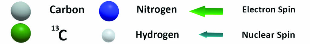
Fluorescent nanodiamonds (FND) are produced from diamond manufactured through high pressure high temperature (HPHT) synthesis. HPHT diamond contains ~100 ppm of nitrogen which is the basis for fluorescent color centers. Since FND is produced by crashing of micron-sized diamond, FND has an irregular shape.
Detonation Nanodiamond Model
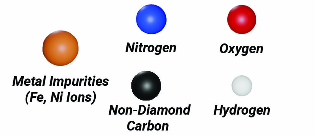
Detonation nanodiamonds (DNDs) are produced from the carbon contained in high-energy explosives. The characteristic size of primary particles is ~ 4-6 nm with very tight size distribution. Raw DNDs are highly agglomerated after synthesis and purification, with average agglomerate sizes of a few hundred nanometers and a wide variety of oxygenated surface functional groups. Large aggregates are separated into individual primary particles (shown on the model). We perform custom surface functionalization: DND enriched with -COOH, -OH, -H, -NH2, and hydrophobic groups are available. Adamas provides monodispersed 5 nm DND with positive or negative zeta potentials in a wide variety of solvents.
Intracellular Targeting of Nanodiamond (ND)
Site-specific intracellular targeting of NDs is a topic of an active research, recently shown by the capacity of fluorescent nanodiamond particles to locally measure free radicals Ref1 or temperature Ref2 in live cells.
Ref1. van der Laan, K. J.; Morita, A.; Perona-Martinez, F. P.; Schirhagl, R., Evaluation of the oxidative stress response of aging yeast cells in response to internalization of fluorescent nanodiamond biosensors. Nanomaterials 2020, 10 (2), 372.
Ref2. Nishimura, Y.; Oshimi, K.; Umehara, Y.; Kumon, Y.; Miyaji, K.; Yukawa, H.; Shikano, Y.; Matsubara, T.; Fujiwara, M.; Baba, Y., Wide-field fluorescent nanodiamond spin measurements toward real-time large-area intracellular thermometry. Sci. Rep. 2021, 11 (1), 1-12
Intracellular Targeting of Nanodiamond (ND)
Step 1— “Intracellular Internalization”: In cellular environments, ND has been shown to be readily taken into cells via clathrin–mediated endocytosis or micropinocytosis.1-2
Step 2— “Endosomal Escape”: After internalization, endosomal escape is necessary to reach targeted structures and has been clearly demonstrated in some works3 The capability of ND to escape endosomes in some cell lines has been attributed to their pointed edges formed from milling of microscopic particles to nanometer sizes that hypothesized to enhance penetration of the endosomal membrane.4-5 Additionally, escape may be facilitated by proper design, for example by functionalization strategies which can encourage escape such as cationic,6 proton-adsorbing,7-8 or membrane-like coatings.9
Step 3— “Targeting Mitochondria”: ND freed from endosomes into the cytoplasm can specifically target sub-cellular organelles such as mitochodria,3, 10 which may be localized by moieties such as antibodies11 (e.g. anti-VDAC2 for mitochondria12) or mitochondrial localizing sequence (MLS).3 Additionally, functionalizing the particle to a positively charged (cationic) surface was shown to induce mitochondrial targeting.10
For colloidal stability of ND conjugated with targeting ligands in biological environments, the addition of a short poly(glycerol) layer has been shown to maintain colloidal stability in water and buffers, while maintain the ability to be linked to targeting moieties by carbodiimide chemistry.13-15
References
2. Sigaeva, A.; Morita, A.; Hemelaar, S. R.; Schirhagl, R., Nanodiamond uptake in colon cancer cells: the influence of direction and trypsin-EDTA treatment. Nanoscale 2019, 11 (37), 17357-17367.
3. Chan, M. S.; Liu, L. S.; Leung, H. M.; Lo, P. K., Cancer-cell-specific mitochondria-targeted drug delivery by dual-ligand-functionalized nanodiamonds circumvent drug resistance. ACS applied materials & interfaces 2017, 9 (13), 11780-11789.
4. Chu, Z.; Zhang, S.; Zhang, B.; Zhang, C.; Fang, C. Y.; Rehor, I.; Cigler, P.; Chang, H. C.; Lin, G.; Liu, R.; Li, Q., Unambiguous observation of shape effects on cellular fate of nanoparticles. Sci. Rep. 2014, 4, 4495.
5. Chu, Z.; Miu, K.; Lung, P.; Zhang, S.; Zhao, S.; Chang, H.-C.; Lin, G.; Li, Q., Rapid endosomal escape of prickly nanodiamonds: implications for gene delivery. Sci. Rep. 2015, 5, 11661.
6. Jarre, G.; Heyer, S.; Memmel, E.; Meinhardt, T.; Krueger, A., Synthesis of nanodiamond derivatives carrying amino functions and quantification by a modified Kaiser test. Journal of Or 2014.
7. Zhang, X.-Q.; Chen, M.; Lam, R.; Xu, X.; Osawa, E.; Ho, D., Polymer-functionalized nanodiamond platforms as vehicles for gene delivery. ACS nano 2009, 3 (9), 2609-2616.
8. Creusat, G.; Rinaldi, A.-S.; Weiss, E.; Elbaghdadi, R.; Remy, J.-S.; Mulherkar, R.; Zuber, G., Proton sponge trick for pH-sensitive disassembly of polyethylenimine-based siRNA delivery systems. Bioconjugate Chem. 2010, 21 (5), 994-1002.
9. Vavra, J.; Rehor, I.; Rendler, T.; Jani, M.; Bednar, J.; Baksh, M. M.; Zappe, A.; Wrachtrup, J.; Cigler, P., Supported Lipid Bilayers on Fluorescent Nanodiamonds: A Structurally Defined and Versatile Coating for Bioapplications. Adv. Funct. Mater. 2018, 28 (45), 1803406.
10. Cheng, Y.; Liu, D.-z.; Zhang, C.-x.; Cui, H.; Liu, M.; Mei, Q.-b.; Lu, Z.-f.; Zhou, S.-y., Mitochondria-targeted antioxidant delivery for precise treatment of myocardial ischemia–reperfusion injury through a multistage continuous targeted strategy. Nanomedicine: Nanotechnology, Biology and Medicine 2019, 16, 236-249.
11. Glancy, B., Visualizing mitochondrial form and function within the cell. Trends Mol. Med. 2020, 26 (1), 58-70.
12. Ramos, L. C.; Rodrigues, F. P.; Biazzotto, J. C.; de Paula Machado, S.; Slep, L. D.; Hamblin, M. R.; da Silva, R. S., Targeting the mitochondrial VDAC in hepatocellular carcinoma using a polyclonal antibody-conjugated to a nitrosyl ruthenium complex. JBIC Journal of Biological Inorganic Chemistry 2018, 23 (6), 903-916.
13. Torelli, M. D.; Rickard, A. G.; Backer, M. V.; Filonov, D. S.; Nunn, N. A.; Kinev, A. V.; Backer, J. M.; Palmer, G. M.; Shenderova, O. A., Targeting fluorescent nanodiamonds to vascular endothelial growth factor receptors in tumor. Bioconjugate Chem. 2019, 30 (3), 604-613.
14. Miller, B. S.; Bezinge, L.; Gliddon, H. D.; Huang, D.; Dold, G.; Gray, E. R.; Heaney, J.; Dobson, P. J.; Nastouli, E.; Morton, J. J., Spin-enhanced nanodiamond biosensing for ultrasensitive diagnostics. Nature 2020, 587 (7835), 588-593.
15. Barton, J.; Gulka, M.; Tarabek, J.; Mindarava, Y.; Wang, Z.; Schimer, J.; Raabova, H.; Bednar, J.; Plenio, M. B.; Jelezko, F.; Nesladek, M.; Cigler, P., Nanoscale Dynamic Readout of a Chemical Redox Process Using Radicals Coupled with Nitrogen-Vacancy Centers in Nanodiamonds. ACS nano 2020.